UPPER BRONCHOSCOPY
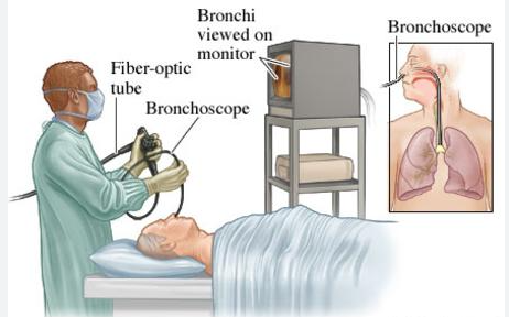
A bronchoscopy involves using a narrow, lighted tube (bronchoscope) to view directly into the lungs’ airways. The mouth or nose is used to insert the bronchoscope. It travels into the airways through the trachea, the windpipe that runs down the throat. The trachea, bronchi, the main airways leading to the lungs, and bronchioles, which are tiny branches of the bronchi, can then be seen by a medical professional.

The Reason for Conducting the Test
To aid in the diagnosis of lung issues by your physician, you might have a bronchoscopy. Your healthcare professional has the ability to examine your airways or obtain a biopsy sample.
Frequently, a bronchoscopy is performed for the following purposes:
An imaging test revealed abnormal lung abnormalities, such as a tumour or growth, lung tissue alterations or scarring, or partial lung collapse.
to biopsy your lungs’ surrounding lymph nodes.
to determine the cause of your bloody cough.To justify dyspnea or low oxygen saturation.
to see if your airway is blocked by anything foreign.
You have been experiencing a cough for almost three months, but the cause is still unknown.
You require a specific kind of diagnostic or have an infection in your major airways (bronchi) and lungs that cannot be identified in any other way.
You breathed in a harmful gas or substance.
to check for the occurrence of lung rejection following a lung transplant.
How the Exam Is Conducted
An instrument called a bronchoscope is used to look inside the lungs and airways. The scope may be pliable or inflexible. Almost invariably, a flexible scope is employed. It is a tube that is roughly two feet (60 centimetres) long and less than half an inch (1 centimetre) wide. Rigid bronchoscopes are utilised in very few instances.
It’s likely that you’ll receive intravenous (IV) medication to assist you relax. Alternatively, if a stiff scope is being utilised, you can be unconscious due to general anaesthesia.
Your lips and throat will be sprayed with an anaesthetic, or numbing agent. In case the bronchoscopy is performed via the nose, numbing jelly will be used to the nostril where the tube is inserted.
Inserting the scope carefully is done. You’ll probably start out coughing from it. As the numbing medication starts to take effect, the coughing will stop.
Saline solution may be injected through the tube by your healthcare professional. In addition to cleaning the lungs, this enables your healthcare professional to take samples of lung tissue, secretions, bacteria, and other things from inside the air sacs. We refer to this phase of the process as a lavage.
Occasionally, very little brushes, needles, or forceps may be inserted via the bronchoscope to remove very small samples of tissue from your lungs (biopsies).
During the surgery, your clinician may also use ultrasonography to evaluate your lungs or implant a stent in your airway. Stents are tiny, tube-shaped medical devices. A painless imaging technique that gives your doctor access to your internal organs is ultrasound.
Occasionally, ultrasonography is used to view the tissues and lymph nodes surrounding your airways, and tiny needles may be introduced to take tissue samples and perform certain diagnostics.
The scope is taken out after the surgery.